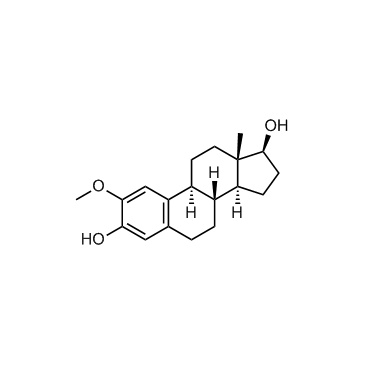
2-Methoxyestradiol
CAS No. : 362-07-2
(Synonyms: 2-ME2; NSC-659853)
| Size | Price | Stock |
|---|---|---|
| 5mg | $25 | In-stock |
| 10mg | $30 | In-stock |
| 50mg | $60 | In-stock |
| 100mg | $70 | In-stock |
| 200 mg | Get quote | |
| 500 mg | Get quote | |
| We match the lowest price on market. | ||
We offer a substantial discount on larger orders, please inquire via [email protected]
or Fax: (86)21-58955996
Inquiry for price and availability only. Please place your order via our email or fax.
| Cat. No. : | HY-12033 |
| M.Wt: | 302.41 |
| Formula: | C19H26O3 |
| Purity: | >98 % |
| Solubility: | H2O : < 0.1 mg/mL (insoluble); DMSO : ≥ 100 mg/mL (330.68 mM) |
2-Methoxyestradiol (2-ME2), an orally active endogenous metabolite of 17β-estradiol (E2), is an apoptosis inducer and an angiogenesis inhibitor with potent antineoplastic activity. 2-Methoxyestradiol also destablize microtubules. 2-Methoxyestradio, also a potent superoxide dismutase (SOD) inhibitor and a ROS-generating agent, induces autophagy in the transformed cell line HEK293 and the cancer cell lines U87 and HeLa[1][2][3][4][5][6].
IC50 & Target:IC50: 1.2 μM (tubulin/microtubule, in living interphase MCF7 cells)[1]
In Vitro: 2-Methoxyestradiol (2-ME) (5-100 μM) inhibits assembly of purified tubulin in a concentration-dependent manner, with maximal inhibition (60%) at 200 μM 2-Methoxyestradiol (2ME2). In living interphase MCF7 cells at the IC50 for mitotic arrest (1.2 μM), 2-Methoxyestradiol significantly suppresses the mean microtubule growth rate, duration and length, and the overall dynamicity, consistent with its effects in vitro, and without any observable depolymerization of microtubules. 2-Methoxyestradiol induces G2-M arrest and apoptosis in many actively dividing cell types while sparing quiescent cells. 2-Methoxyestradiol binds to tubulin at or near the colchicine site, it inhibits microtubule assembly, and high concentrations have been shown to depolymerize microtubules in cells[1].
2-Methoxyestradiol (2-ME) decreases the HIF-1α and HIF-2α nuclear staining in cells cultured under hypoxia. 2-Methoxyestradiol is an anti-angiogenic, anti-proliferative and pro-apoptotic agent that suppresses HIF-1α protein levels and its transcriptional activity. A significant decrease in the growth rate is found in the 10 μM 2-Methoxyestradiol-treated A549 cells in comparison with the DMSO-treated cells (66.2±7.2 and 101.2±2.3%, respectively; p=0.04) at 96 h. A significant increase in apoptosis is observed in cells treated with 10 μM 2-Methoxyestradiol in a normoxic condition in comparison with cells under lower O2 concentration (5.8±0.2%; p=0.003)[2].
In Vivo: To investigate the effect of 2-Methoxyestradiol (2-ME2) on uveitis development, C57BL/6 mice are randomly assigned into two groups and immunized with IRBP peptide. 2ME2 group starts 2-Methoxyestradiol (15 mg/kg) intraperitoneally from day 0 to day 13 while control group is given with vehicle. The disease score of 2-Methoxyestradiol (2ME2) group is 0.30±0.30, significantly lower than that of control group 2.09±0.28 (p<0.05), each group containing 5 mice[3].
Treatment with 2-Methoxyestradiol (60-600 mg/kg/d) results in a dose-dependent inhibition of tumor growth. The percentage of cells with strong pimonidazole-positive staining (+++) is significantly decreased in the 2-Methoxyestradiol-treated group (36.0% for 60 mg/kg/d and 0% for 200 and 600 mg/kg/d) compare with the vehicle-treated group (86.5%). This may be attributed to the dramatic inhibition of tumor growth in a dose-dependent manner following 2-Methoxyestradiol treatment[4].
Lorem ipsum dolor sit amet, consectetur adipisicing elit. Autem earum hic iste maiores, nam neque rem suscipit. Adipisci consequatur error exercitationem fugit ipsam optio qui, quibusdam repellendus sed vero! Debitis.
Inquiry Information- Product Name:
- 2-Methoxyestradiol
- Cat. No.:
- HY-12033
- Quantity:
Your information is safe with us.