
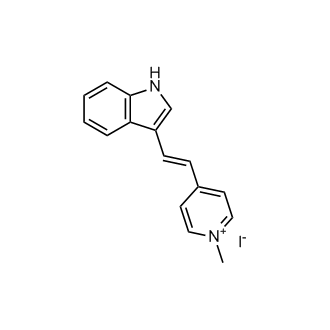
F16
CAS No. : 36098-33-6
| Size | Price | Stock |
|---|---|---|
| 5mg | $38 | In-stock |
| 10mg | $60 | In-stock |
| 25mg | $120 | In-stock |
| 50mg | $216 | In-stock |
| 100 mg | Get quote | |
| 200 mg | Get quote | |
| We match the lowest price on market. | ||
We offer a substantial discount on larger orders, please inquire via [email protected]
or Fax: (86)21-58955996
Inquiry for price and availability only. Please place your order via our email or fax.
| Cat. No. : | HY-100395 |
| M.Wt: | 362.21 |
| Formula: | C16H15IN2 |
| Purity: | >98 % |
| Solubility: | Ethanol : 1 mg/mL (ultrasonic);DMSO : ≥ 31 mg/mL |
F16 is a mitochondrial permeability transition pore (PTP) modulator with an EC50 of 30 μM in mice. F16 promotes PTP opening, accumulates in cancer cell mitochondria, and induces mitochondrial depolarization, swelling, cristae disruption, outer membrane rupture, ATP depletion, reactive oxygen species (ROS) production, cytochrome c release and mitochondrial uncoupling. F16 induces cell cycle arrest at the G1 (occasionally G2) phase, reduces the phosphotyrosine content of Neu, as well as the levels of phosphorylated PKB/Akt and phosphorylated MAP kinase, downregulates the protein expression levels of Neu and PKB, triggers apoptosis in cells with moderate Bcl-2 expression, and induces necrosis in cells with overexpressed Bcl-2. F16 can be used in research related to breast cancer and gastric cancer[1][2][3].
In Vitro:F16 (100 nM-15 μM; 36-40 h) selectively inhibits proliferation of EpH4-A6 neu-overexpressing mouse mammary epithelial cells, with partial inhibition observed at concentrations as low as 100 nM[1].
F16 (3 μM; 3-7 days) inhibits proliferation of neu-, v-Ha-ras-, and β-catenin-initiated mouse mammary tumor cell lines and most human breast cancer cell lines[1].
F16 (3 μM; 36 h-3 days) induces cell cycle arrest (primarily G1 arrest) in EpH4-A6 neu-overexpressing mouse mammary epithelial cells and sensitive mouse and human breast tumor cell lines[1].
F16 (24 h) induces S phase cell cycle arrest in SGC-7901 cells after 24 h of incubation[3].
F16 (3 μM; 15-72 h) induces apoptosis in EpH4-A6 neu-overexpressing mouse mammary epithelial cells, characterized by oligonucleosomal DNA fragmentation, Annexin V positivity, and cytochrome c release, but does not induce apoptosis in EpH4-EV control cells[1].
F16 (0.3-3 μM; 48 h) induces oligonucleosomal DNA fragmentation (apoptosis) in apoptosis-sensitive EpH4-A6 and SMF cells, but not in apoptosis-resistant EpH4, Bcl-2-overexpressing EpH4-A6.C13, EpH4-A6.C18, SMF-Bcl-2, or NF324-1C cells[2].
F16 (3 μM; 18-36 h) induces cytochrome c release (an apoptotic marker) in apoptosis-sensitive EpH4-A6 cells within 18 h, but not in apoptosis-resistant Bcl-2-overexpressing EpH4-A6.C13 and EpH4-A6.C18 cells, where delayed cytochrome c localization changes are associated with mitochondrial breakdown rather than apoptosis[2].
F16 (3 μM; 24 h) induces caspase-3 activation (an apoptotic marker) in apoptosis-sensitive EpH4-A6 and SMF cells, but not in apoptosis-resistant Bcl-2-overexpressing EpH4-A6.C13, EpH4-A6.C18, SMF-Bcl-2, or NF324-1C cells[2].
F16 (Various concentrations; 24 h) induces apoptosis in SGC-7901 cells in a concentration-dependent manner after 24 h of incubation[3].
F16 (3 μM; 2-24 h) accumulates selectively in mitochondria of sensitive mouse and human mammary tumor/transformed epithelial cell lines, but not in resistant normal or tumor cell lines[1].
F16 (3 μM; 1 h) accumulation in mitochondria is driven and retained by the mitochondrial transmembrane potential (ΔΨm), and lack of accumulation in resistant cell lines is not mediated by MDR-1 or MRP-1 pumps[1].
F16 (1 μM; 1 h) does not alter accumulation in EpH4-A6 cells due to Bcl-2 overexpression, with F16-sensitive cell lines showing higher mitochondrial accumulation than F16-resistant cell lines[2].
F16 (3-50 μM; 15-72 h) compromises mitochondrial structure and function in sensitive cells, causing swelling, ATP depletion, superoxide production, permeability transition pore opening, and altered respiration, leading to cytotoxicity[1].
F16 (3 μM; 24-48 h) downregulates Neu-dependent signaling in EpH4-A6 neu-overexpressing mouse mammary epithelial cells, reducing phosphorylation of Neu, PKB, and MAPK, and decreasing Neu and PKB protein levels[1].
F16 (3 μM; 24-96 h) induces a time-dependent reduction in cell number in apoptosis-sensitive EpH4-A6 and SMF cells, as well as apoptosis-resistant Bcl-2-overexpressing EpH4-A6.C13, EpH4-A6.C18, SMF-Bcl-2, and NF324-1C cells, with a 3 μM concentration causing up to 32-fold reduction in cell number over 96 h in EpH4-A6 cells[2].
F16 (3 μM; 24-48 h) induces apoptosis in EpH4-A6 cells and necrosis in Bcl-2-overexpressing EpH4-A6.C13 and EpH4-A6.C18 cells; caspase inhibition reduces F16-induced death in EpH4-A6 cells, while buffering ATP levels and neutralizing superoxide reduces necrotic death in Bcl-2-overexpressing cells[2].
F16 (3 μM; 48 h) induces apoptotic morphological changes in EpH4-A6 cells and necrotic morphological changes in Bcl-2-overexpressing EpH4-A6.C13 cells after 48 h of incubation[2].
F16 (3 μM; 15-48 h) induces a time-dependent decrease in cellular ATP levels in both apoptosis-sensitive EpH4-A6 and SMF cells, and apoptosis-resistant Bcl-2-overexpressing EpH4-A6.C13, EpH4-A6.C18, SMF-Bcl-2, and NF324-1C cells, with a 50-60% reduction in ATP levels observed after treatment in SMF-derived and NF324-1C cells[2].
F16 (48 h) has its cytotoxicity on SGC-7901 cells almost completely reversed by ATP pre-treatment 0.5 h before F16, indicating that decreased intracellular ATP availability is a major factor in F16-mediated cytotoxicity[3].
F16 (3 μM; 15-72 h) induces a time-dependent increase in superoxide anion levels in apoptosis-sensitive EpH4-A6 cells and apoptosis-resistant Bcl-2-overexpressing EpH4-A6.C13 and EpH4-A6.C18 cells, but not in F16-resistant EpH4-EV cells[2].
F16 (48 h) potently inhibits proliferation of SGC-7901 and MCF-7 cancer cells with over 10-fold selectivity for SGC-7901 cells over non-tumor GES-1 cells[3].
F16 (48 h) increases intracellular ROS levels in SGC-7901 cells after 48 h of incubation, with partial reversal by pre-treatment with reductants[3].
F16 (3 µM; 24 h) selectively accumulates in the mitochondria of SGC-7901 cells after 24 h of incubation at 3 µM[3].
F16 (48 h) reduces intracellular ATP levels in SGC-7901 cells after 48 h of incubation, with no additional effect at higher concentrations[3].
F16 (10 µM; higher concentrations) induces dose-dependent structural damage to isolated rat liver mitochondria, including swelling at 10 µM and membrane rupture at higher concentrations[3].
F16 enhances H+ and K+ permeabilization of the inner membrane of isolated rat liver mitochondria in a concentration-dependent manner[3].
F16 immediately increases the membrane fluidity of isolated rat liver mitochondria, as measured by reduced fluorescence anisotropy of HP-labeled mitochondria[3].
F16 (75 µM) increases state 4 oxygen consumption rate of isolated rat liver mitochondria in a dose-dependent manner, with a 3-fold increase at 75 µM, indicating an uncoupling effect[3].
F16 (20 µM) increases the metabolic thermogenic output of isolated rat liver mitochondria at 20 µM, consistent with an uncoupling effect on oxidative phosphorylation[3].
F16 causes a concentration-dependent decrease in mitochondrial membrane potential (ΔΨm) in isolated rat liver mitochondria, which is mediated by mitochondrial permeability transition (MPT)[3].
F16 (1 h) induces concentration-dependent release of cytochrome c from isolated rat liver mitochondria after 1 h of incubation at 4 °C[3].
In Vivo:F16 (20 mg/kg; i.p.) retards the growth of EpH4-A6-derived subcutaneous breast tumors in nude mice[1].
Lorem ipsum dolor sit amet, consectetur adipisicing elit. Autem earum hic iste maiores, nam neque rem suscipit. Adipisci consequatur error exercitationem fugit ipsam optio qui, quibusdam repellendus sed vero! Debitis.
Inquiry Information- Product Name:
- F16
- Cat. No.:
- HY-100395
- Quantity:
Your information is safe with us.